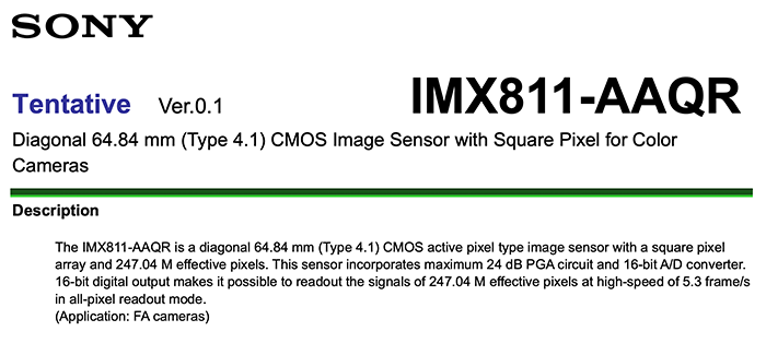
Could that be the new Full Frame 16 megapixel Monochrome Sony sensor?

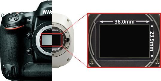
Many months ago I reported how Sony was developing a Full Frame monochrome sensor. Interesting enough Nikon just announced a new DS-Qi2 monochrome camera for microscope use. It has a 16.2 Megapixel Full Frame monochrome sensor “that features high pixel density, high sensitivity and low noise, making it an excellent choice for applications in quantitative fluorescence imaging.“. As you know “most” of the Nikon FF sensors are made by Sony so is that one a Sony FF too?
I hope we will get such kind of sensor on a real RX(?) camera soon!
Here are the sensor details:
Key Features
High Sensitivity
Detects even faint fluorescent signals
7.3μm pixels, high quantum efficiency, and very low read noise allow the DS-Qi2 to read in even faint fluorescent signals.

Excellent Linearity
Reliable quantitative analysis made possible
With a linearity error of ±1%, the DS-Qi2 is a superb tool for measuring intensities in fluorescence samples, including timebases intensity measurement and radiometric measurements.
High Frame Rate
Fast focusing, even with fluorescent images
With a high-sensitivity CMOS sensor and USB 3.0-based data transfer, the DS-Qi2 enables high-speed live imaging and image capture at up to 45 fps (1636×1088 pixels).
Low Noise
Acquires dim fluorescent signals with ultra-low noise
2.2 electrons read noise coupled with a large full-well capacity allows the acquisition of fluorescence images with very little noise and very high dynamic range.


LLC-PK1 cells expressing GFP-EB3 tubulin with low noise. Large linear full well capacity allows acquiring both the brightest and dimmest areas in a single capture.
Sample courtesy of: Michael Davidson, National High Magnetic Field Laboratory, Florida State University
Time-lapse Photography
Fluorescent time-lapse imaging through integration with NIS-Elements software

With a large field of view and pixel density, and low noise, the DS-Qi2 is ideal for time-resolved imaging applications.

Time-lapse images (every 1 second) of LLC-PK1 cells with GFP-EB3 tubulin. Each image represents the maximum intensity projection of the time-lapse, allowing visualization of the end-binding protein located on the microtubule plus-ends, and allowing tracing of the microtubule path.
DS-Qi2 captures an extremely large field of view, but still represents very fine details as demonstrated in this cropped time-lapse sequence from a large FOV image.
Objective: CFI Plan Apochromat λ 60x oil / NA: 1.4
Sample courtesy of: Michael Davidson, National High Magnetic Field Laboratory, Florida State University